
This project is a door that will open up a new era of digital pathology for me.

Due to the Covid pandemic and some procedural waiting times, it took nine months to get here, but finally, I am in Norway. I was able to come with the special permission applied for me. Since the special permission period was short, I packed my things and said goodbye to everyone in a short time. Although I was fully vaccinated, I had to stay at the hotel as quarantine and later in my house for ten days when I arrived in Norway. This period was a bit difficult and strange, but I tried to make it fun and effective by reading a novel and working on my project.
On my first day, I met a lot of people at the hospital and in the laboratory. Luckily, everybody is so friendly and helpful. They helped me a lot to get used to everything and answered all my questions. I realized how different it is to work remotely and on-site. Once again, I understood the importance of working on-site and communicating with people. I am responsible for two master students from my first day. We meet regularly, and I try to teach my students the histopathology of breast cancer. I taught them how to evaluate mitosis, PPH3 and Ki67 in the pathological slides. We also learned from our colleague the CISH (chromogenic in situ hybridization) procedure for staining miRNA (microRNA) and how we evaluate it. Getting knowledge on miRNA is essential for my project; since previous studies show the presence of specific miRNAs correlating with TNBCs (Triple Negative Breast Cancer) and the presence of TILs (Tumor infiltrating lymphocytes).
miRNAs are a class of small non–protein-coding RNA molecules of ~9–24 base pairs in length, involved in RNA silencing and post-transcriptional regulation of gene expression. These are one of the largest groups of translational regulators in human cells. They have shown higher expression in many different tumor types. Two types of miRNA-18a and miRNA-18b are expressed in the stroma of TNBCs with a high degree of inflammation and potentially associated with macrophages. Figure 1 shows an example of specific staining for miRNA-18a/b as a dark blue color in the stroma of a TNBC which performed by CISH. CISH is based mainly on the same immunogenic principles as immunohistochemistry.
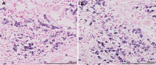 Figure 1: A. miRNA-18a by CISH, B. miRNA-18b by CISH
These results suggest the expression of miRNA-18a/b are correlated with systemic immunological response in ER-negative tumors. Therefore, I examine the relevant and corresponding cell types and importance for the prognosis of miRNAs in TNBC.
During these months, half of the Clarify project ESRs are also in Norway. We communicate with each other and work together. Communicating with other PhD candidates and working together gives me the opportunity to comprehend other projects and get a better perspective on the final goal of the CLARIFY. Besides scientific work, it feels so good to have entertaining plans with other project members (Figure 2). I think having fun plans with my team members might also improve my professional efficiency at work and job satisfaction. Although I have been enjoying working on my project under the supervision of Emiel when I was in Turkey, working in Emiels` team on-site, have taught me a lot so far. In the next post, I will explain more about the prognostic factors of TNBC.
Figure 2: Preikestolen (Pulpit rock)
Umay kiraz – ESR11.