
Tuning deep learning architectures for detecting artifacts in bladder cancer digital slides.

Computers are smarter at many tasks, but their intelligence comes from the data they experience. Digitization of pathological glass slides is the first step in this puzzle. The routine of preparing histopathological glass slides introduces different unintentional artifacts and variations due to decalcification, manual dying, and scanning hardware. Digital Pathology (DP) is a term used for the workflow of acquiring, managing, and interpreting a Whole Slide Image (WSI). WSI, in general, contains an ample amount of information for developing algorithms for automated analysis, such as quantification, detection, and characterization of molecular subtypes. Apart from challenges like anomalies in the data that may encourage wrong decisions in computers. Automation of pathological assessments is critical for mitigating the projected rise of cancer rates in the near future. Therefore, it is essential to understand the complexity of this workflow and improve it further to aid existing infrastructure.
An image analysis pipeline can influence the algorithm’s overall performance in the computer by ensuring the fidelity of relevant information. Artifacts such as folds, bubbles, damage, and blur deleverage learning of relevant features by adding irrelevant morphological features that do not carry any histological information. Thus, the use of deep learning models for artifact detection can undermine the risk of unreliable prediction and help develop efficient CAD systems. We are testing several existing architectures by measuring the impact of different processing techniques on the end task. Once these models are tuned for identifying certain image instances, they can evaluate the presence of anomalies in downstream diagnosis tasks. We are observing the pleasant outcome of models. The results seem promising for their potential use in preprocessing pipeline, but there is also room to improve.
Currently, I am attending a month-long secondment at Stavanger University Hospital to look further into the pathological perspective of diagnostical relevance in Whole Slide Images. This collaboration will help us understand the characteristics of three cancer types: Non-Muscle Invasive Bladder Cancer, Triple Negative Breast Cancer, and Spitzoid Melanocytic Lesions. I am pretty sure that understating the biology of cancer may help navigate through other digital pathology challenges.
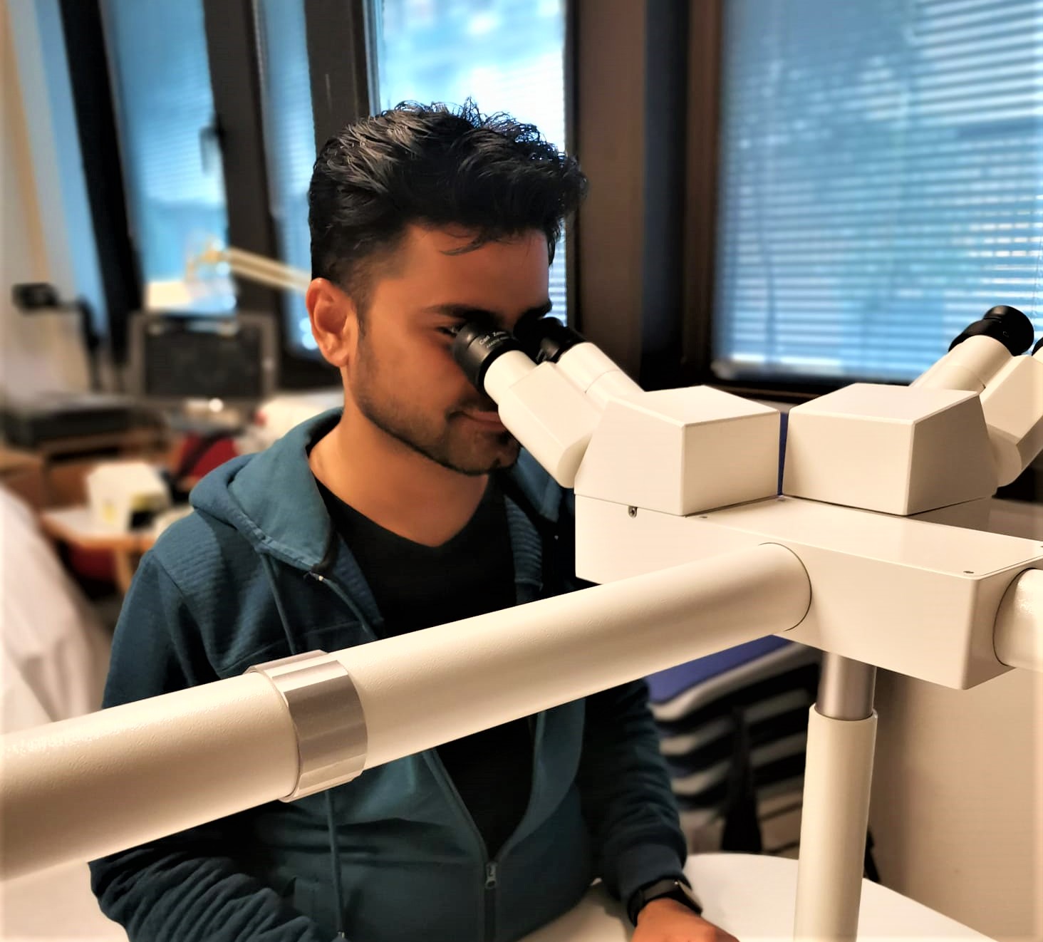
Experiencing conventional microscopy to look into histological digital slide
Neel Kanwal – ESR4