
A monumental progress in deep learning for healthcare technology is not possible without knowledge of digital pathology.
ESR1 is a computer science research position in CLARIFY working on Semantic interoperability of digital pathology data. Her interests are data discovery and research asset search. ESR4 is also a computer science research position in CLARIFY, but ESR4 focuses on preprocessing and region of interest for different cancer types. His research project is designed for several outcomes such as segmentation models and anonymization algorithms. ESR10 is a biomedical engineer. He is focusing on bladder cancer. In the CLARIFY project, he aims to improve the diagnosis and prognosis of high-risk non-muscle invasive bladder cancer (HR-NMIBC) patients by using artificial intelligence (AI) and digital pathology. In those contexts, in order to complement their training, they participated together in a virtual secondment at Stavanger University Hospital (SUH).
This virtual secondment was a two-month training aimed to enhance pathological understanding of this multidisciplinary project. It was comprised of series of lectures, sessions with a uropathologist, recorded seminars, and open discussions. This secondment can be divided into three major segments based on quantitative pathology, digital pathology workflow, and digital pathology diagnosis.
Quantitative pathology (QP) can provide important diagnostic and prognostic information that might improve cancer patients` treatment possibilities. It provides awareness that changes can be detected quantitatively. QP is more about making subjective measurements objective and improving reproducibility. Digital pathology enables better quality control and calibration for each laboratory. The major of moving to digital slides is standardization. We have constrained our focus to H&E only because we are going to annotate images in our database which are in this staining only. Meanwhile, digital pathology (DP) enables many functionalities that would have been impossible or tricky for traditional pathology, such as annotation, automatic measurement, proof of diagnostics, online clinical meetings, consultations, quantitative research, etc. The disadvantages of DP also exist, e.g., extra cost for scanners and data storage, the large size of whole slide images. Inevitably, the digitalization of glass slides has posed challenges on information technologies to support efficient storage, processing and sharing of high-volume and large-sized digital images. But at the same time, we can see opportunities to improve digital pathology workflow and eventually achieve a faster and more accurate diagnosis.
During the secondment, the pathologist introduced different histopathological features and structures, such as different tissue types, papillary structure, cancer cell invasion, cell polarity, pleomorphism, etc., and demonstrated how to recognize different features from the slide images with real-world examples. In particular, the uropathologist starts examining a digital slide at 10x to observe the region of interest. This region of interest varies from the prospect of analysis, but it is the main area with cellular structures. Urothelium is generally a tissue type with an abundance of cell nuclei. A magnification of 40x is generally suitable to observe cellular polarity. Also, the uropathologist annotated the regions of interest for image analysis to elaborate on the annotation of a whole slide image (WSI).
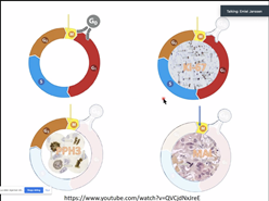
Figure 1: Screenshot from one of the online seminars
The DICOM standard for digital pathology has come a long way since its inception. Immunohistology quantification has an excellent impact on the automatic evaluation of digital images. Moreover, the secondment gave a prime insight into the pathological analysis of NMIBC, and biomarkers used for breast cancer. We attended lectures related to cancer diagnosis based on cellular features. It was a good insight to understand the multidisciplinary nature of the project. The parameters to evaluate cancer grading and variants of histological features to understand the impact of treatment. Stavanger University Hospital has the biggest pathological center in the region, and they are doing great work for cancer mitigation with every possible technological advancement. Despite all the inconvenience in a virtual form, the lectures are informative and interesting. We learned many pathological terms and skills.
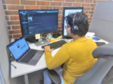
Figure 2: Neel Kanwal participating in the secondment at SUH.
Na Li, Neel Kanwal and Farbod Khoraminia – ESR 1, ESR4 & ESR10