
“The controversy induced by Spitz tumors represents a never-ending challenge for pathologists, so we believe that using artificial intelligence may be the solution to make a step forward in tackling this problem.”

In the last post, we presented the clinical context of Spitz tumors. As promised, today we will talk about how we, Andrés and Laëtitia (ESR12 and ESR7), plan to tackle this challenging problem!
The study of Spitz tumors is two-fold, for which we will work hand in hand. The aim is to use Artificial Intelligence (AI) to identify the tumor and integrate the morphological data to be able to render a specific diagnosis based on the individualized prediction of the metastatic risk of this ambiguous tumor, allowing the best therapeutic approach.
Therefore, the analysis of Spitz tumors will have both a medical (Andrés) and an engineering (Laëtitia) approach, leading to two different Ph.D. subjects. Laëtitia will focus on the AI algorithms, while Andrés will work on the clinical aspect.
More precisely, the objectives of each thesis are described below:
A. Medical focus (Andrés, ESR12)
-
- Digitize the biopsies and annotate the whole-slide images of spitzoid melanocytic tumors;
- Validate deep learning algorithms for the diagnosis and prognosis of ambiguous spitzoid melanocytic tumors;
- Study the histopathological patterns and clues of spitzoid melanocytic tumors, together with immunohistochemical, genetic and clinical information.
B. Engineering focus (Laëtitia, ESR7)
-
- Develop innovating deep learning-based algorithms for the classification of the input images into healthy or pathological regions for spitzoid melanocytic tumors;
- Detect the malignant potential of clinically controversial spitzoid melanocytic cases;
- Obtain the corresponding features for the characterization of spitzoid melanocytic lesions;
- Adapt algorithms and the approach depending on ESR12’s feedback.
Figure 1 sums up the main objectives and how we will combine our work to carry out this research.
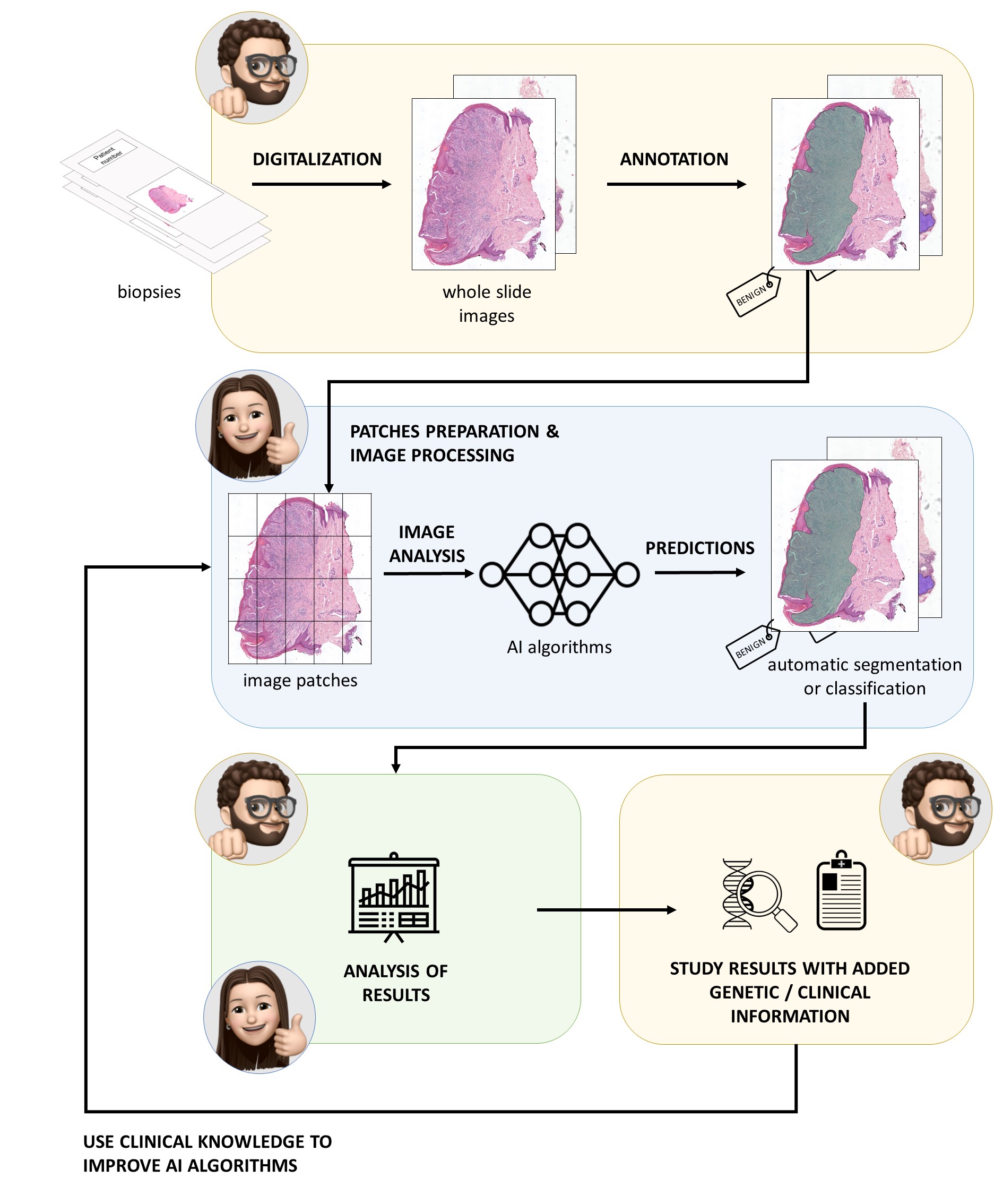
Figure 1. Main steps of the two Ph.D. projects of Andrés (yellow blocks) and Laëtitia (blue blocks), working hand in hand through the whole process.
ESR7 Laëtitia and ESR12 Andrés.