
The quick automated detection of hemorrhage in CT scans can help save lives – our multiple instance learning algorithm gives accurate prediction without the need of slice level annotations.

Intracranial hemorrhage (ICH) is a life-threatening emergency with high rates of mortality and morbidity. Rapid and early detection of ICH is essential because nearly 30% of the life loss happens in the first 24hours.The diagnosis of ICH is performed using CT brain scans (see Figure 1) that consist of multiple slices (typically ~35 per scan). This process requires the fast and accurate examination of a radiologist. In order to prompt the optimal treatment to patients in short time, computer-aided diagnosis (CAD) is being designed to establish a better triaging protocol.
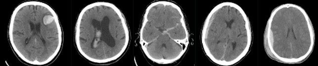
Figure 1: Five slices of a CT brain scan for hemorrhage detection.
Artificial Intelligence (AI) has shown impressive results in medical image classification but usually requires huge amounts of annotated data. Existing approaches for ICH detection often depend on time-consuming slice level annotations of radiologists to train the AI model.
We developed an approach of Multiple Instance Learning (MIL) that is able to reach competitive results using only scan-level annotations which are easier to obtain. From the MIL perspective, the slices are called ‘instances’ and the complete CT scans are called ‘bags’. During training, only the bag-level labels are available (in our case the diagnosis of the complete CT scan). The proposed model combines a convolutional neural network with attention mechanism with variational Gaussian processes resulting in a two phase training procedure. We experimentally show that the model provides accurate and fast slice- and scan-level predictions of ICH. For more information, please read our preprint https://www.biorxiv.org/content/10.1101/2021.07.01.450539v1.
The article is the result of a successful collaboration of the University of Granada with the ESR-8, Arne Schmidt, supervised by Professor Rafael Molina and the Northwestern University with PhD candidate Yunan Wu and Professor Aggelos Katsaggelos who is an External Advisory Board member of CLARIFY. The MICCAI conference is one of the worlds best conferences of medical image analysis and will will be held from September 27th to October 1st 2021 as a virtual event.
For future research we are eager to discover similar multiple instance learning approaches with probabilistic models for histopathological images. Allthough the cancer classification with Whole Slide Images (WSIs) is a different area in medicine it can also be formulated as a MIL problem: Each WSI represents a bag and all image patches are the instances. If the WSI diagnosis (bag label) is available we can apply similar methods as in the presented paper.
Thank you for your attention and keep reading our blog if you are interested in the papers yet to come!
Arne Schmidt – ESR8