
Being able to be a part of a dermatopathology service and learn about the practical aspect of cutaneous lesions’ diagnosis has nothing to do with what you can read in books.

I had the chance to be the first ESR of the CLARIFY network to do an on-site secondment this year. For two months, April and May, I was at the INCLIVA foundation where Andrés (ESR12) is doing his Ph.D. under the tutelage of Carlos Monteagudo.
This secondment brought me so much! Not only did I learn about dermatopathology but I also got to work hand in hand with Andrés.
Before the secondment, my knowledge of dermatopathology was almost none, except for the literature of the field I had read and the theory I had studied. But being able to be a part of a dermatopathology service and learn about the practical aspect of cutaneous lesions’ diagnosis has nothing to do with what you can read in books and the theory. Like when you learn a new language, the best way to learn is to immerse yourself!
During these two months, I could attend the daily sign-outs of biopsies with expert dermatopathologists. Thanks to these daily sessions in which pathologists analyse the biopsies with microscopes to make a diagnosis, I could become more familiar with the specific terminology of the field. I learnt to recognise the different layers of the skin and their components (sebaceous glands, vessels, subcutaneous fat tissue, etc.) as well as the different types of cells, such as melanocytes, lymphocytes, etc. Being able to recognise healthy skin is an essential step to be able to detect abnormalities and lesions.
I remember how it all seemed a tremendous amount of new information in the beginning, when I did not understand much during the daily sign-out sessions. However, little by little —and thanks to the help and explanations of Carlos and Andrés—, this information started to make more sense to me, and I actually had a great time trying to diagnose different types of lesions!
The most recurrent lesion type was usually basal cell carcinoma, which can have many morphologic subtypes, i.e. superficial, nodular, micro nodular, infiltrative. There were also a lot of melanocytic proliferation lesions, such as nevus —standard ones but also dysplastic or congenital ones, among others. As for spitzoid lesions, the ones that represent the main focus of Andrés’ (ESR12) thesis project as well as mine, these are more uncommon: I think we saw less than 4 or 5 during the daily sign-outs, in the two months I spent at the INCLIVA!
Despite not seeing many “new” Spitz tumours, learning about the different types of lesions and characteristics of some of them, additionally to healthy skin features, helped me understand better and thus become able to recognise more efficiently the different characteristics of Spitz tumours. Lesions are never as clear in practice as the photo examples we see in books or articles! Besides, Andrés explained to me in more detail the different features of spitzoid lesions on the whole-slide images on MicroDraw, the web platform pathologists use for annotating in CLARIFY.
In short, I learnt a lot thanks to Carlos and Andrés, but also to the whole team! The acquired knowledge in dermatopathology will definitely be helpful to interact more efficiently with experts in the field using the adapted vocabulary. But, most of all, it will make a difference at the moment of analysing the results of the artificial intelligence models developed, but also to better define the next steps and approaches of the project.
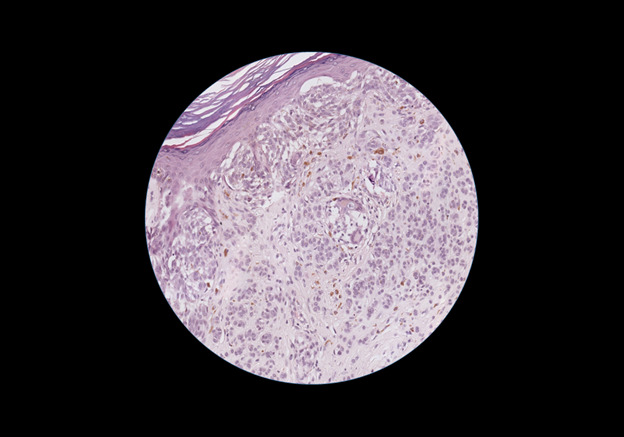
Visualisation of a spitzoid tumour on the microscope.
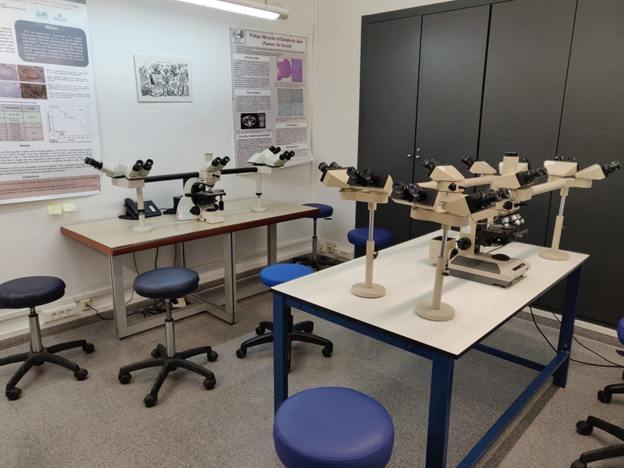
The room with microscopes where we did the daily sign-outs. There are some microscopes where we can be up to 10 people looking at a biopsy at the same time!
Laëtitia – ESR7.