
Multidisciplinary research will be only possible if you collaborate with your colleagues with diverse expertise.

You might think annotating the histopathological slides efficiently for training or testing deep learning algorithms needs only pathological knowledge; If so, I am going to explain why you should adjust the way you are thinking. In this post, I am explaining the learning process of how to annotate whole slide images in our study and why I believe it is vital to learn from both pathologists and computer scientists.
As one of the PhD candidates in the CLARIFY project, a part of my work is to provide our computer scientist collaborators with annotated whole slide images. So, I have been learning how to annotate whole slide images within our dataset to answer our research questions properly. We have to teach the computer different parts in the images: urothelium layer, lamina propria, cancerous vs normal, artefacts, etc. Obviously, it would help if you gained a proper level of knowledge in pathology to recognize different areas in a whole slide image. Therefore I had several training sessions with Prof. van Leenders (uropathologist) at Erasmus MC to learn the basics of uropathology. Following these sessions, I annotated several slides, and I realized that still, some challenges exist which were not pathology related but computer science-related.
Whether you want to train a child or an algorithm, you should talk to them in a language they understand. So, for training an algorithm, you should learn how to speak in the computer language, and in the case of annotation, you should take into account several points:
- It would help if you tried to annotate with high specificity. For example, if you are annotating stroma, you should avoid including any other tissue types.
- The more annotation you get, the better the algorithm output you get. Although the annotation process is very time consuming, you should try to annotate as much as possible. Therefore, you should find a balance between detailed annotation and the number of annotated slides.
- Annotate some areas that a pathologist never annotate in daily slide revision. For example, for detecting cancerous vs non-cancerous regions, you should annotate normal urothelium as well. In contrast, pathologists don’t annotate non-cancerous regions in regular slide revision due to a lack of prognostic importance.
- We are annotating based on our research questions. Sometimes you should annotate some areas for pre-processing of whole slide images like artefacts and different tissue types. These are some areas that pathologists never annotate on a daily slide revision as well.
These are several essential points for annotation that don’t come from pathology but from the way you should train the computer. I am so happy to have annotation review meetings at my secondment in Stavanger. These meetings are with Two pathologists (Mr V. Kvikstad and Ms U. Kiraz) and a computer scientist (Mr S. Fuster), and we discuss annotations on our slides weekly (Figure 1). Then, I annotate the whole slide images according to the points we discuss in each session.
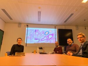
Figure 1: Meeting with two pathologists and a computer scientist on reviewing annotations
Therefore, multidisciplinary research will be only possible if you collaborate with your colleagues with diverse expertise. Delineating whole slide images for training algorithms would be effective when different experts comment on the delineations. These multidisciplinary discussions have taught me how to annotate the slides so far effectively.
Farbod Khoraminia – ESR10